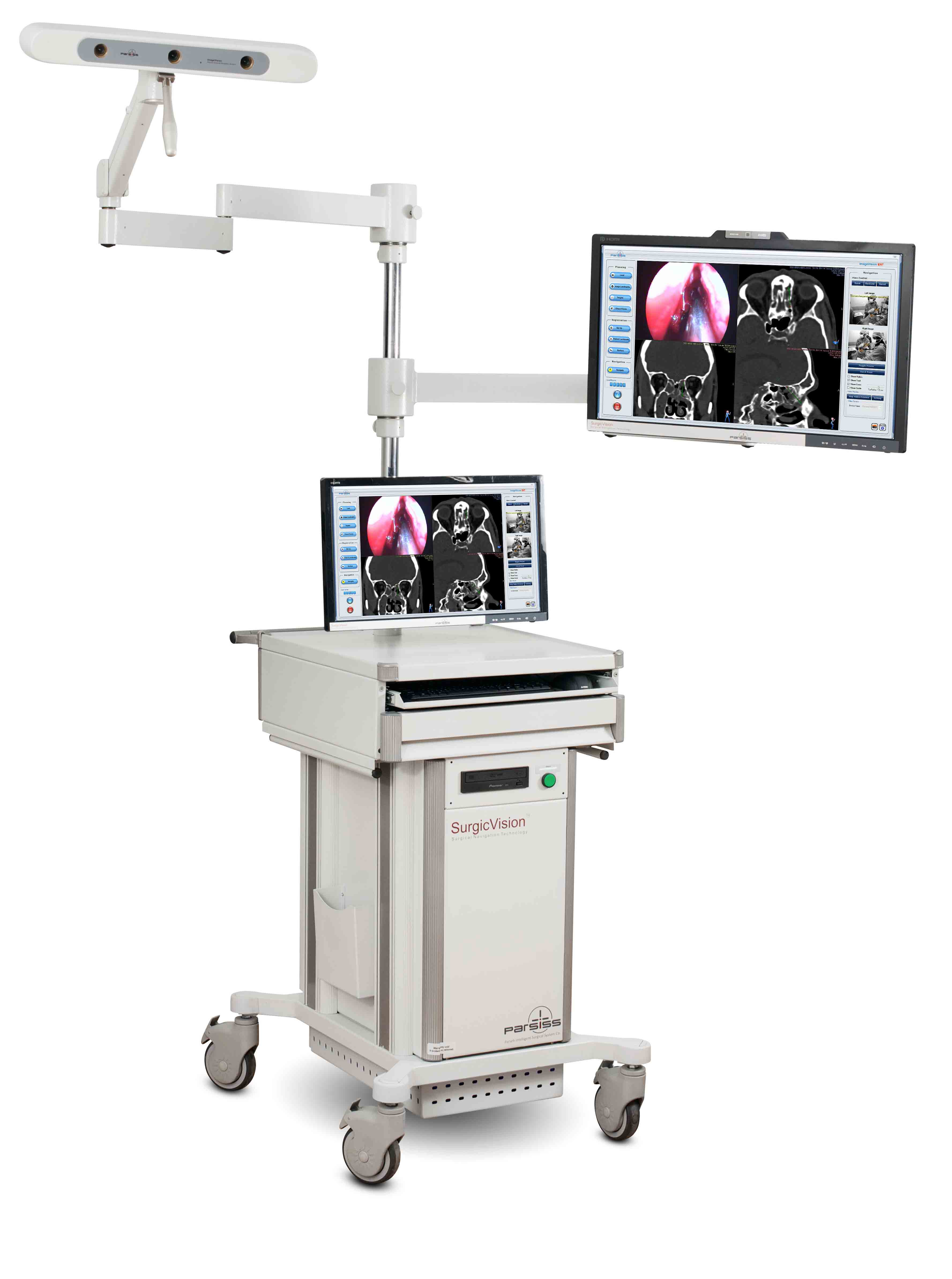
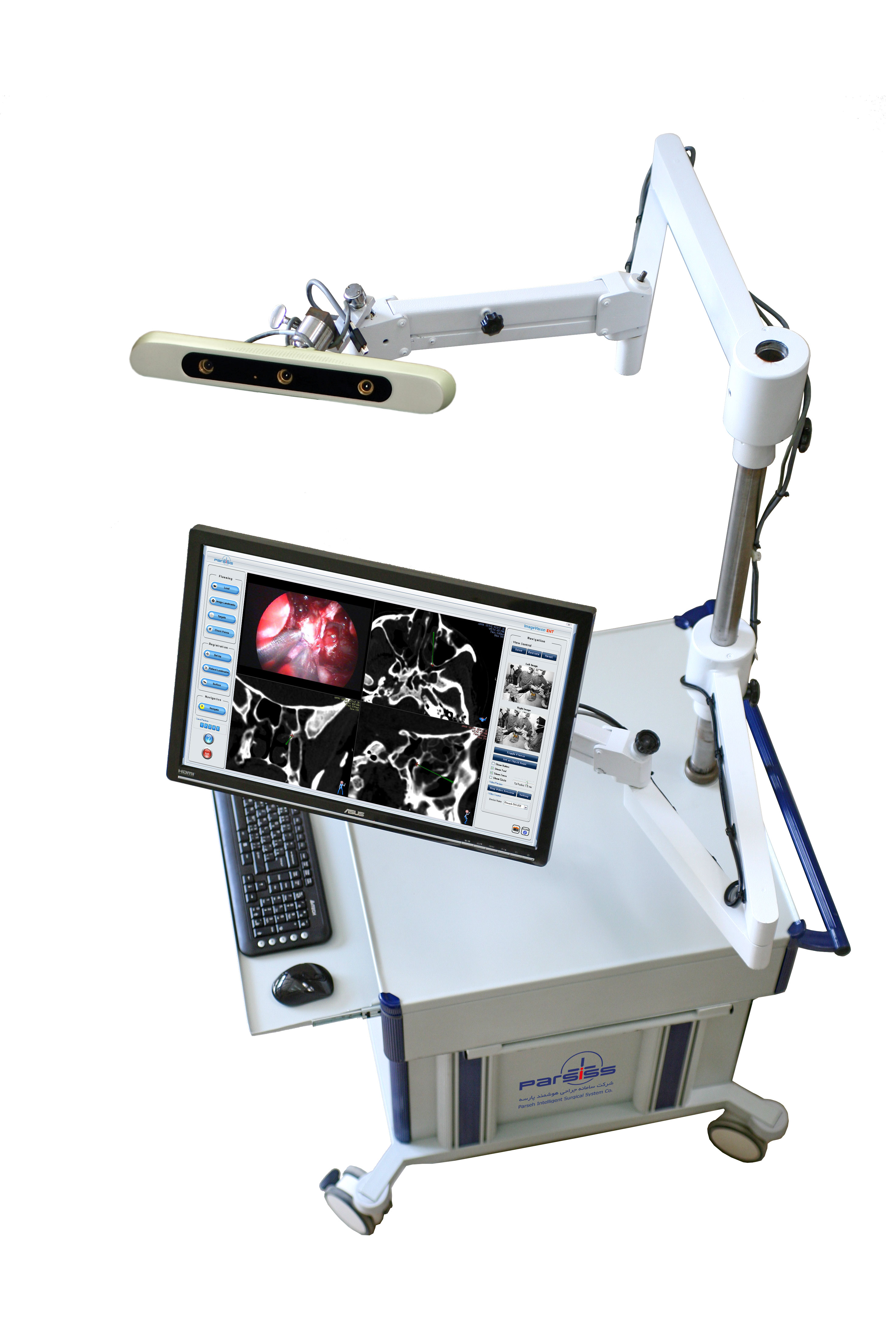
IV2
IV2 is a high performance surgical navigation system for ENT surgeries. Using advanced technology and high precision optical camera it offers surgeons more accurate and reliable navigation during surgery.
IV2 has fast, user friendly and robust software which provides simple and minimal hand interaction by more visualized and automatic procedures. This comprehensive software offers advanced facilities from pre-operative planning to post-operative documentation.
System supports wide range of data inputs such as CT scan, MRI and fMRI images and fuses them precisely. It has full integration with operation room devices such as microscope, endoscope and different imaging devices.
Ear/Nose/Throat applications
As with cranial surgery, extreme finesse is required to navigate the many small passages and cavities involved in ENT surgery. The navigation system can be used for:
- Sinus surgery
- Cyst and polyp removal
- Optic nerve decompression
- Frontal mucocele
- Skull base surgeries
Import & Manage Image Dataset
|
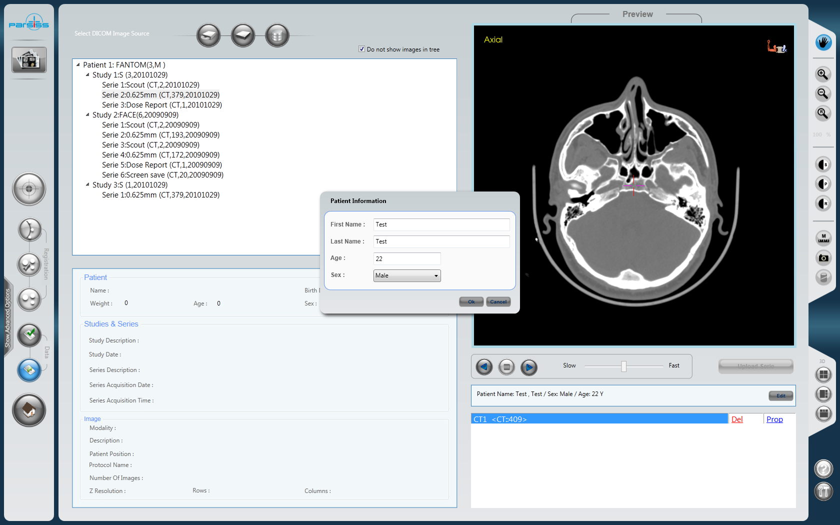
Visualization
|
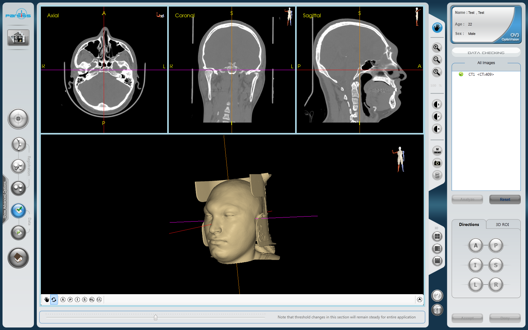
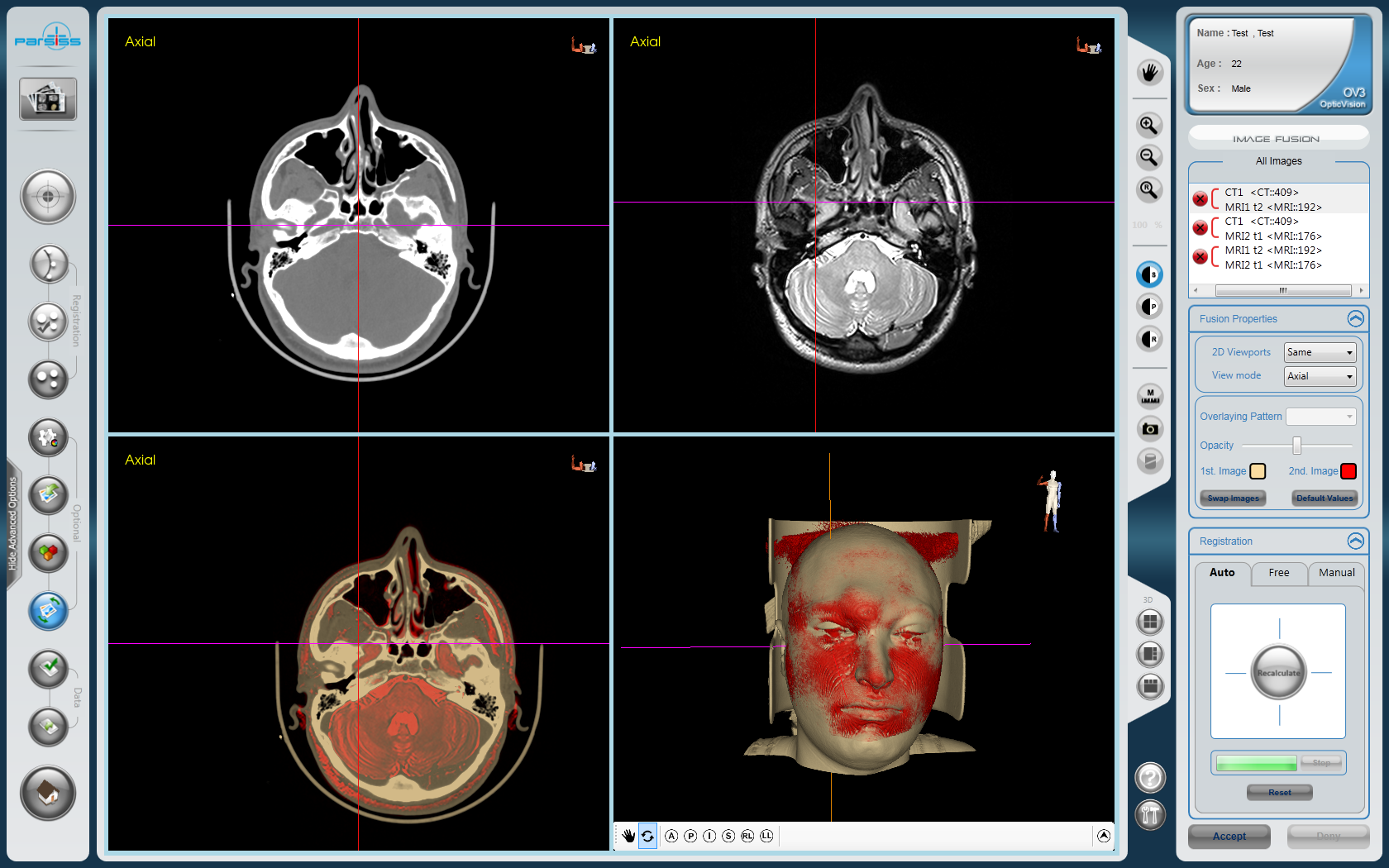
Image Fusion
- Automatic fusion of images from different image modalities, e.g., CT and MR
- Accurate and fast fusion, using advanced algorithms
- Fusion refinement using manually selected landmarks
- Fusion of image datasets with different voxel size
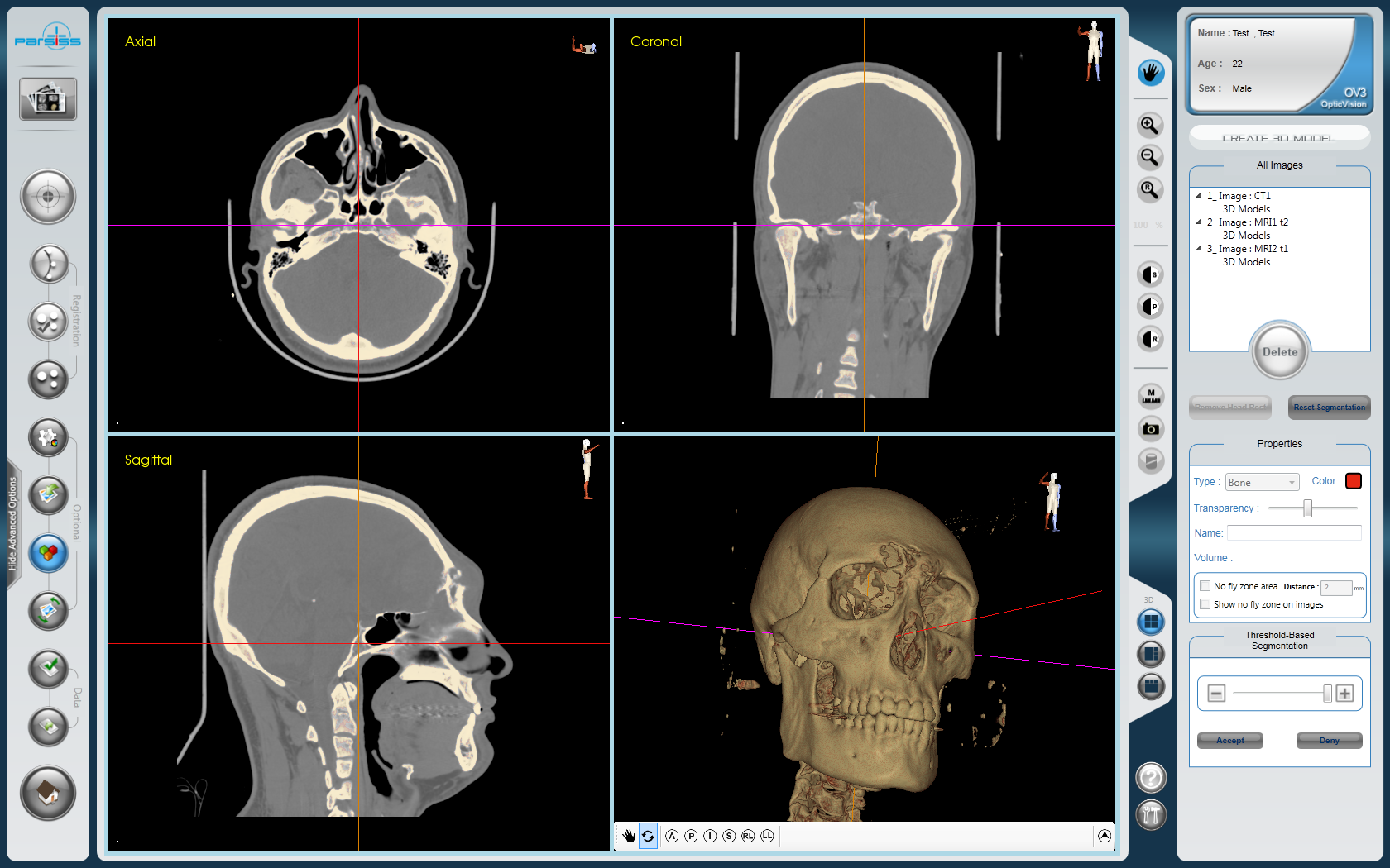
Segmentation & 3D Reconstruction
- Automatic segmentation of bone and skin from CT images
- Easy semi-automatic segmentation of the selected regions
- Display segmented organs with predefined tissue colors
- Color and opacity adjustment of segmented regions
- Capability to specify the VOI (Volume Of Interest) for segmentation
- Fast 3D model reconstruction
- Display sections of 3D models in standard 2D views
- Capability to define critical anatomical regions to caution during the surgery
- Automatic volume calculation of segmented objects
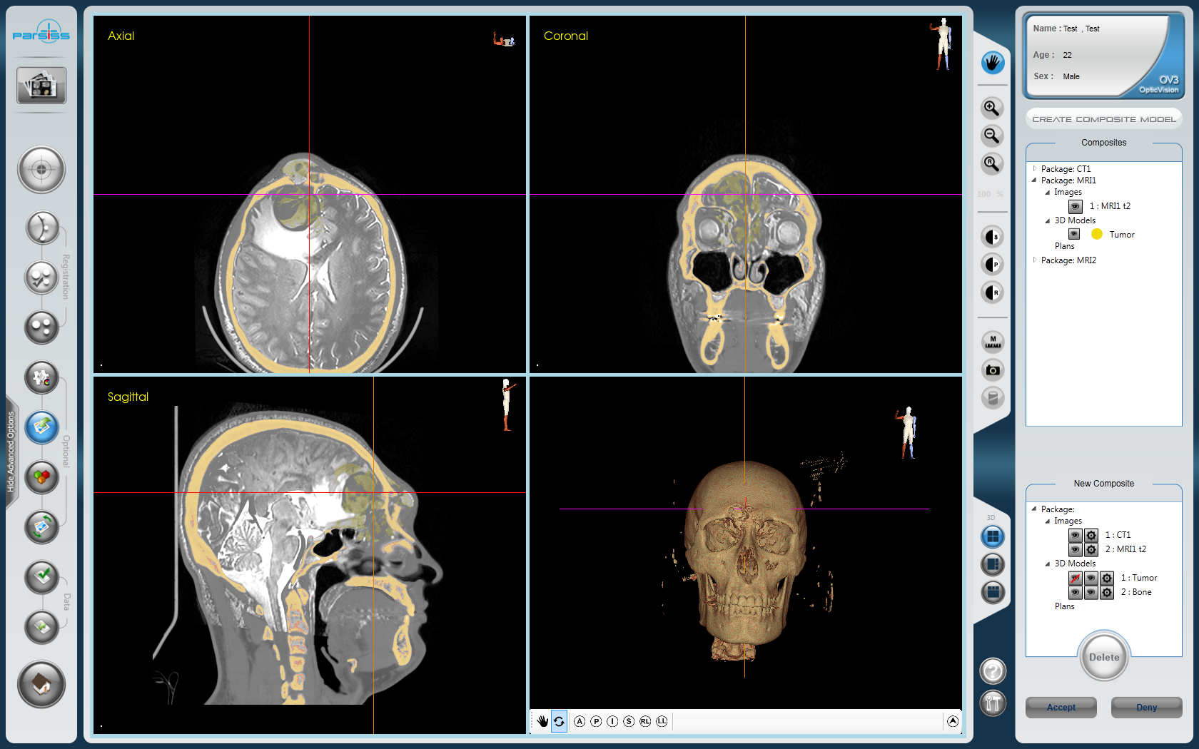
Composite Modeling
- Fast and easy overlay of fused data and 3D models from different modalities
- Individual opacity adjustment of composite model’s section
- Automatic validity check of the composite model
- Automatic correction of the composite model based on updated fusion results
- Capability to create multiple composite models
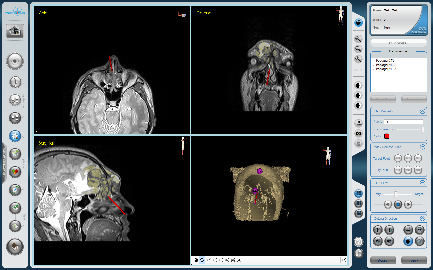
Surgical Planning
- Preoperative simulation of surgical procedure in virtual environment
- Capability to set and modify target and entry points on 2D images or 3D model
- Capability to adjust visualization parameters of surgical path, e.g., color and opacity
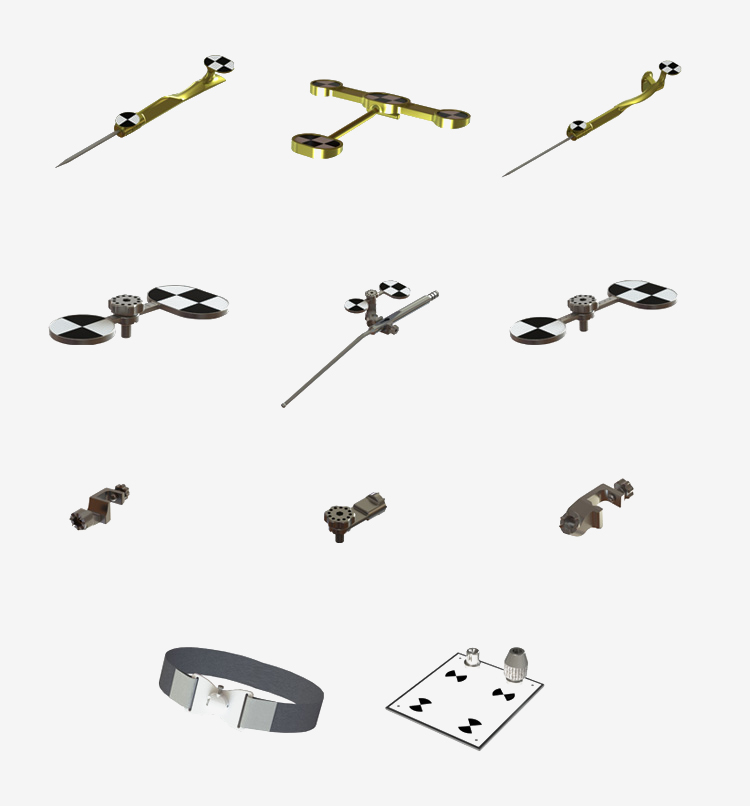
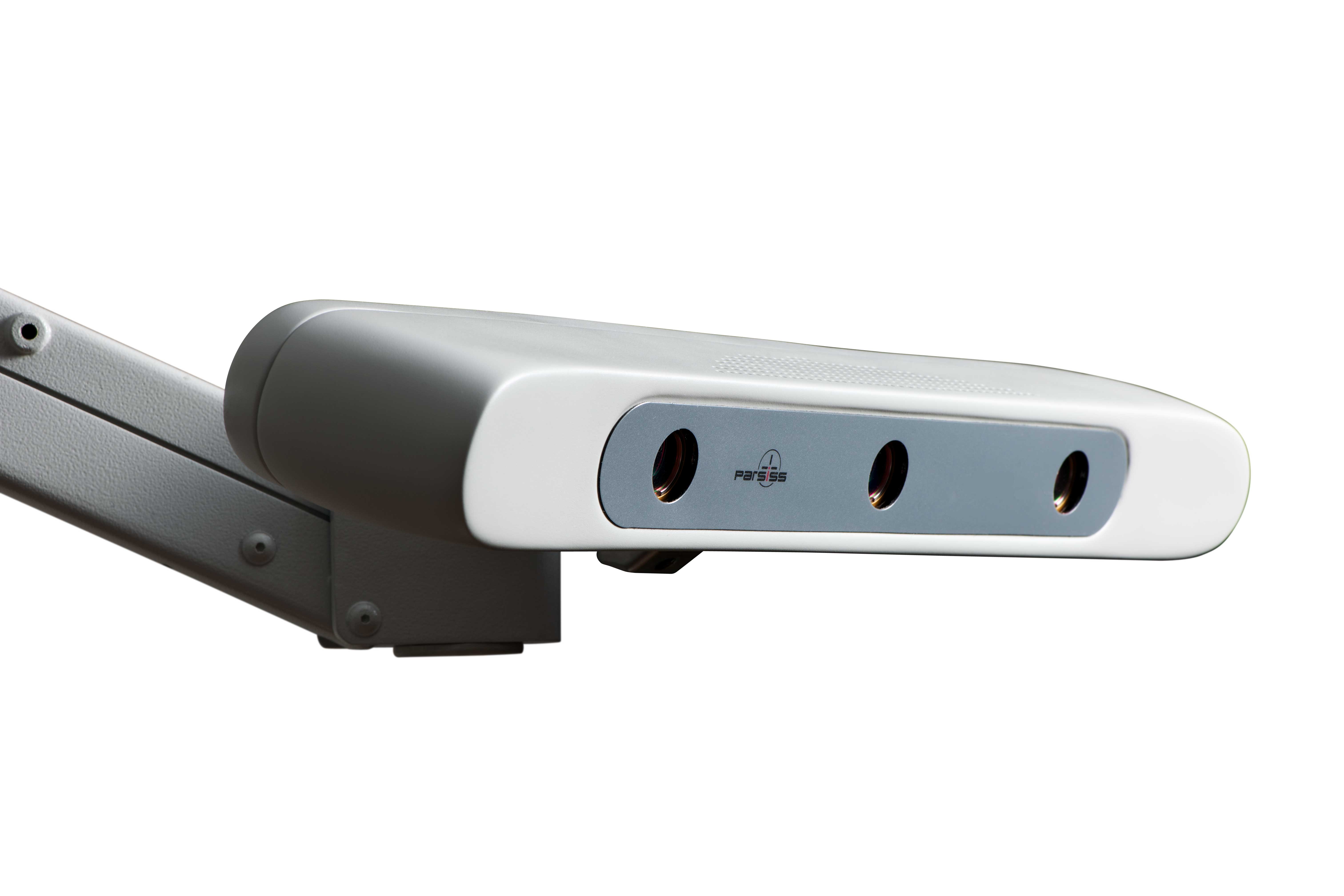
Tracking Device & Tool Support
- Extensive field of view
- Support active and passive surgical instruments
- Automatic detection and validation of surgical instruments
- Easy calibration and visual accuracy check of surgical instruments
- Capability to deactivate/reactivate instruments in the system
- Handy and multiface instruments to facilitate surgical maneuver
- Optimal adjustment of tracker’s field of view using laser pointer
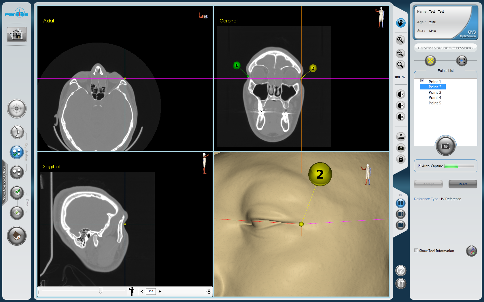
Registration
- Minimal invasive registration
- Suggestion of predefined anatomical landmarks
- Flexible landmark selection in 2D and 3D views
- Display landmark registration error in 2D and 3D model for registration refinement
- Automatic landmark capturing
- Automatic evaluation of the new captured landmarks
- Surface registration to achieve higher registration accuracy
- Edit the surface points to refine the surface registration
- Inform final registration error
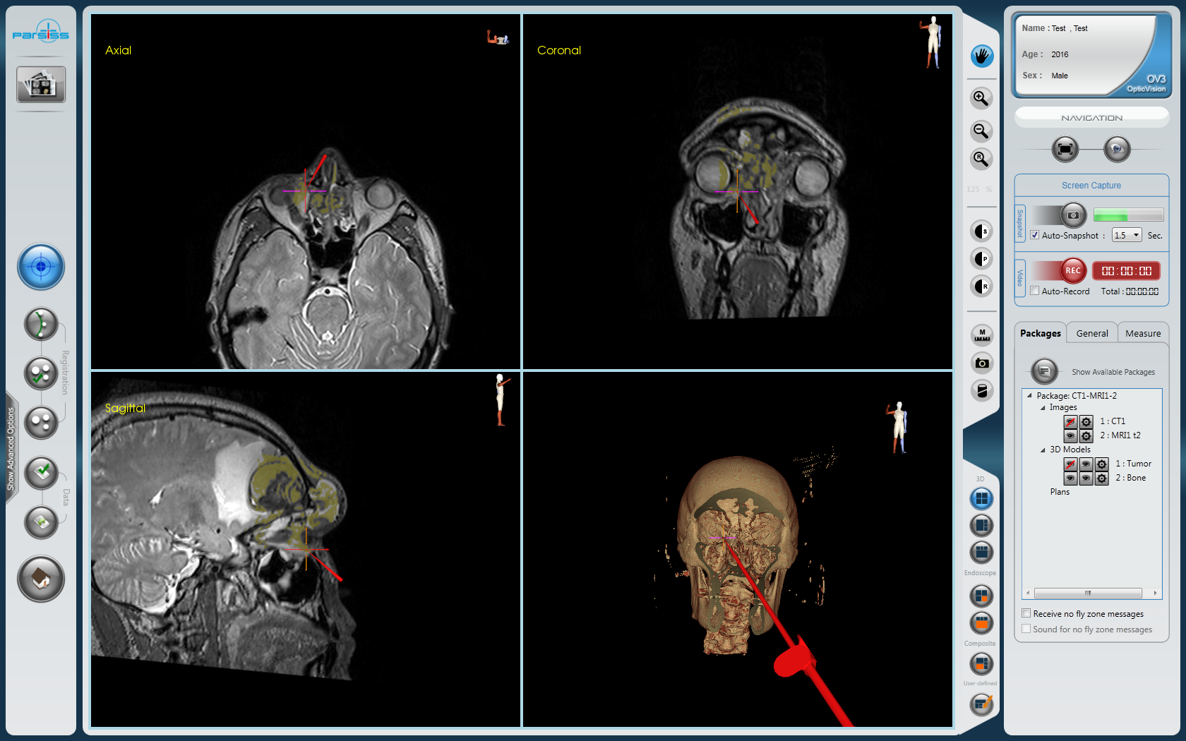
Navigation
- Display the surgical tool’s orientation and tip position on the 2D images and 3D model
- Capability to define check points for intraoperative verification of registration accuracy
- Dynamic cutting 3D of the model at current position of the tooltip
- Detect and compensate camera and patient displacement
- Import and display video images during navigation ,e.g., microscopic and endoscopic images
- Capability to freeze images in order to review, measure and other operations
- Display fused images in 2D and 3D views
- Audio-visual alarm when the tracker misses the reference or tools
- Audio-visual alarm when the tooltip approaches to predefine to critical area
- Intraoperative surgical simulation using virtual tip
- Real time display of distance between predefined target points and tooltip
- Auto-measurement of distance from tooltip to arbitrary points
- 3D distance measurement
- Capability to back review the medical images during navigation
- Automatically switch to full-screen mode
- Auto-snapshot surgeon’s view of interest
Other Features
- Virtual key and foot switch to activate predefined system tasks
- Auto saving and possibility to recover based on the last saved data and setting
- Quick help to guide the user in each step
- UPS support to continue working in the case of power failure
Accessories